Journal of Biology and Medicine
Comparative Evaluation of Cross-Linked Collagen Membrane with and without Silk Fibroin around Immediate Dental Implants-Randomized Controlled Trial
1Postgraduate student, Department of Periodontology, Krishnadevaraya Dental College, Bengaluru. 562157, INDIA
2Reader, Department of Periodontology, Krishnadevaraya Dental College, Bengaluru. 562157, INDIA
3Prof. and Head, Department of Periodontology, Krishnadevaraya Dental College, Bengaluru. 562157, INDIA
Author and article information
Cite this as
Naik JT, et al. Comparative Evaluation of Cross-Linked Collagen Membrane with and without Silk Fibroin around Immediate Dental Implants-Randomized Controlled Trial. J Biol Med . 2026; 10(1): 1-8. Available from: 10.17352/jbm.000049
Copyright License
© 2026 Naik JT, et al. This is an open-access article distributed under the terms of the Creative Commons Attribution License, which permits unrestricted use, distribution, and reproduction in any medium, provided the original author and source are credited.Background and Objectives: Immediate dental implants placed at the time of extraction help preserve alveolar bone, maintain soft tissue architecture, and reduce treatment time. However, compromised bone and soft tissue deficiencies may affect implant outcomes, requiring regenerative approaches. Silk fibroin derived from Bombyx mori has gained attention as a biomaterial because of its favourable mechanical properties and biocompatibility. This study aimed to compare cross-linked bovine collagen membranes with and without silk fibroin around immediate dental implants for soft tissue regeneration.
Materials and methods: Twenty healthy individuals were randomised into two groups following non-surgical periodontal therapy. Group I (Control) received a Cross-linked collagen membrane following immediate implant placement (n = 10), while Group II (Test) received a cross-linked collagen membrane with silk fibroin (n = 10). Immediate implant healing was assessed at the end of the 10th day, 1 month, and 3 Months postoperatively. Gingival tissue thickness (GTT) and keratinised tissue width (KTW) were evaluated at 1 month and 3Months. Bone assessment was performed at baseline and at 3 months post-intervention using Radiovisiography.
Results: Silk fibroin membrane demonstrated significant gingival tissue thickness and enhanced keratinised tissue width with improved wound healing, and greater peri-implant bone stability compared to collagen membrane alone.
Conclusion: Collagen membranes incorporated with silk fibroin may enhance soft tissue regeneration and peri-implant bone stability around immediate implant sites.
Clinical Relevance: Functional composite membrane (Silk fibroin +Collagen) for Soft and Hard tissue augmentation around immediate implants provides greater peri-implant stability.
Replacement of missing teeth is essential to restore function and esthetics, with Dental implants being a predictable and widely accepted option. Implant placement may be immediate or delayed, with immediate placement performed at the time of extraction offering advantages such as reduced treatment time, preservation of tissue architecture, and improved patient comfort. However, in the case where immediate implants are placed in periodontally compromised sites presents clinical challenges like soft tissue deficiencies, reduced keratinised tissue, bone loss, and difficulty in achieving primary stability. The quality and quantity of peri-implant soft tissue, particularly gingival thickness and keratinised tissue width, are critical for long-term success, influencing esthetics, plaque control, and resistance to inflammation. The thickness of the gingival and bone tissue influences treatment outcomes, especially in compromised cases due to changes in blood supply to the underlying bone and vulnerability to resorption [1]. Regenerative procedures are performed before or during implant placement to overcome the loss of volume, thereby restoring alveolar bone [2].
Soft tissue augmentation procedures with autogenous grafts are considered the gold standard but are limited by donor site morbidity. Collagen membranes are widely used due to their biocompatibility and have the capacity to induce mineralisation by active invasion of osteoblastic cells, leading to complete ossification of the membrane [3]. Till date, there is only one case report by Smidt et al (2019), who explored the use of a collagen scaffold as a core material for guided bone regeneration in the case of a horizontal defect around an implant and reported that the procedure is simpler to perform with a favourable outcome [4]. Silk fibroin, a biocompatible protein with superior strength and controlled degradation, offers potential benefits. In preclinical models by Lee et al. (2024), silk fibroin (SF) membranes have demonstrated bone regeneration comparable to commercially used collagen membranes (e.g., Bio-Gide®) in calvarial defects, with similar volumes of new bone formation at 8 weeks, suggesting SF is a viable alternative GBR membrane material with lower pathogen and cost concerns than collagen membranes. In the present study, we have prepared a composite membrane (collagen–silk fibroin membrane) to enhance stability and healing. This study evaluates its effectiveness in improving peri-implant soft tissue and bone outcomes around immediate implants [5].
Materials and methods
Twenty patients were recruited from the outpatient department of periodontology, Krishnadevaraya College of Dental Science and Hospital, satisfying the inclusion and exclusion criteria. A computer-generated randomisation method was followed for grouping. A sealed envelope was opened before the immediate implant placement for allocation into 2 groups. This clinical trial was conducted in accordance with the revised CONSORT 2013 guidelines. The present study was approved by the Institutional Review Board and Ethical Committee and has been registered in “Clinical Trials.gov” with identity number NCT06552065.
Preparation
The Collagen membrane and Composite membrane were prepared by incorporating silk fibroin in a cross-linked collagen membrane at Fibroheal Wound Care Pvt Limited. Newtown Yelahanka Bengaluru 560064.
Type I collagen membrane
Collagen membrane was fabricated according to Jiang S 2021. Bovine tendon was used to extract type I collagen using the acid swelling–pepsin digestion process. Briefly, the fresh bovine tendon fat was removed. Then, the residues were pulverised and soaked in 0.05 mol/l Tris buffer for 24 h. The precipitate was gathered. After that, the pepsin-containing acetic acid solution was added, and the supernatant was gathered. For salting out, a 3.5 mol/l NaCl solution was added. For five days, dialysis was performed with deionised water at 40C. Culture dishes were utilised to determine the collagen solution’s concentration. The culture dish was weighed as M0 and was dry and clean. The culture dish was filled with the recommended amount of collagen solution, and the weight was noted as M1. Next, the culture dish was placed in a 600C constant temperature drying oven until the collagen was fully dried. The weight was recorded as M2. The concentration of collagen solution was calculated according to the formula: (M2 - M0)/ (M1 - M0) 100%. The above experiment was repeated three times, and the mean value was taken [6].
Composite membrane (Collagen and Silk fibroin)
Preparation of hydrogel scaffold: Collagen gel was added to fibroin solution to create fibroin/collagen blends in water. In this study, the weight percentage of collagen in the dried blend hydrogel is fixed at 20%. In the fibroin solution, the collagen gel dissolved when heated to 400 C with gentle stirring.
At 600 c, all of the collagen gel denatured and dissolved in the fibroin solution, forming a translucent fibroin/collagen solution in the process. The collagen and fibroin aqueous solution was concentrated at 500–600 c, while being gently stirred until the fibroin concentration reached 3.4–5%. After the mixed solution had cooled to room temperature, different components of EDC (Table I) were added, and the pH value was adjusted to 4–5 by adding 1 mol/L sodium hydroxide. The fibroin/collagen hydrogel was immediately freeze-dried using a lyophilizer. The freeze-dried scaffolds were cleaned with 1 M NaCl and 0.1 M Na2HPO4 (pH 9.1), in that order. The matrices were then freeze-dried once more after being cleaned with distilled water. To create fibroin/collagen scaffolds devoid of EDC, the fibroin/collagen solution was immediately freeze-dried using a lyophilizer once the pH value was brought to 4-5 [7].
Sterilisation and storage
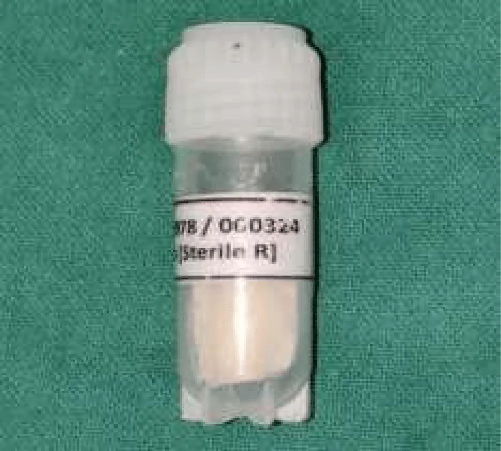
The membranes were prepared and were in 15 × 30 mm dimensions. Following this, it was sterilized using UV radiation and provided in sealed Eppendorf packages. Packages were opened only at the site of intervention.
Case selection and grouping
Patients with compromised teeth, willing for extraction and immediate dental implant placement, were randomised following Phase I therapy. Group I: 10 Patients received cross-linked collagen membrane following immediate implant placement. Group II: 10 Patients received cross-linked collagen membrane with silk fibroin following immediate dental implant placement.
Inclusion criteria: Systemically healthy patients aged between 18 and 55 years undergoing immediate implant placement with gingival thickness ≤1 mm were included in the study. Patients diagnosed with Stage II or Stage III and Grade B periodontitis were considered. Implant site characteristics included assessment of gingival tissue thickness(GTT), keratinised tissue width (KTW) and Radiographic assessment was carried out using Radiovisiography (RVG) with a grid for evaluation.
Exclusion criteria: Patients with known allergies to collagen or silk products were excluded from the study. Individuals with medical conditions contraindicating surgical intervention, known smokers and alcoholics, and patients presenting with acute infections at the extraction site, such as abscesses, were also excluded. Additionally, patients with a history of radiotherapy or chemotherapy were not considered for inclusion in the study.
Clinical parameters
Gingival Tissue thickness (GTT) and Keratinised Tissue Width (KTW)
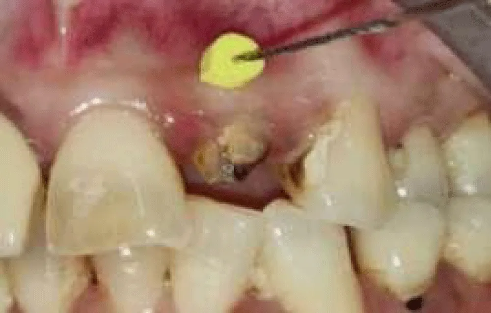
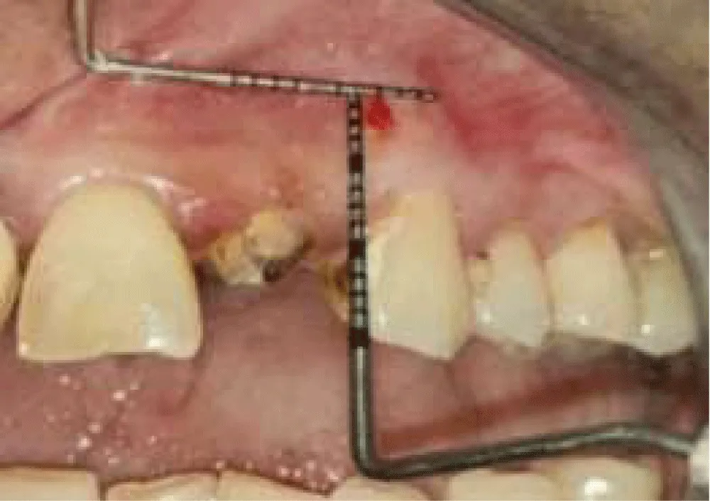
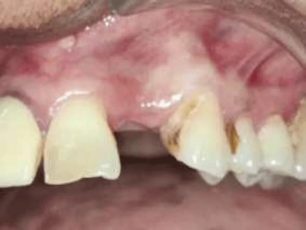
Gingival tissue thickness is the distance of the bucco-lingual width at the designated site. Assessment was done at a point 1.5 mm below the gingival margin of the adjacent tooth at the site of implant placement. Local anaesthesia was administered before the procedure, and an Endodontic K-file with silicone disc stop was pierced in the tissue, and this was followed by measurement using UNC-15 probe. (Figure 1) Keratinised Tissue Width is the distance measured from the gingival margin to the mucogingival junction at the allocated site. Apico-coronal width of keratinised tissue is measured as the distance from the mucogingival junction to the gingival margin, with the mucogingival junction determined using Schiller’s Potassium Iodide Solution. (Figure 2).
Wound healing index
Wound healing index was assessed using Landry Turnbull and Howley Healing Index in that tissue colour, bleeding on palpation, granulation tissue, incision margin, and suppuration were evaluated. (Figure 6).
Radiographic assessment with Radiovisiography (RVG)
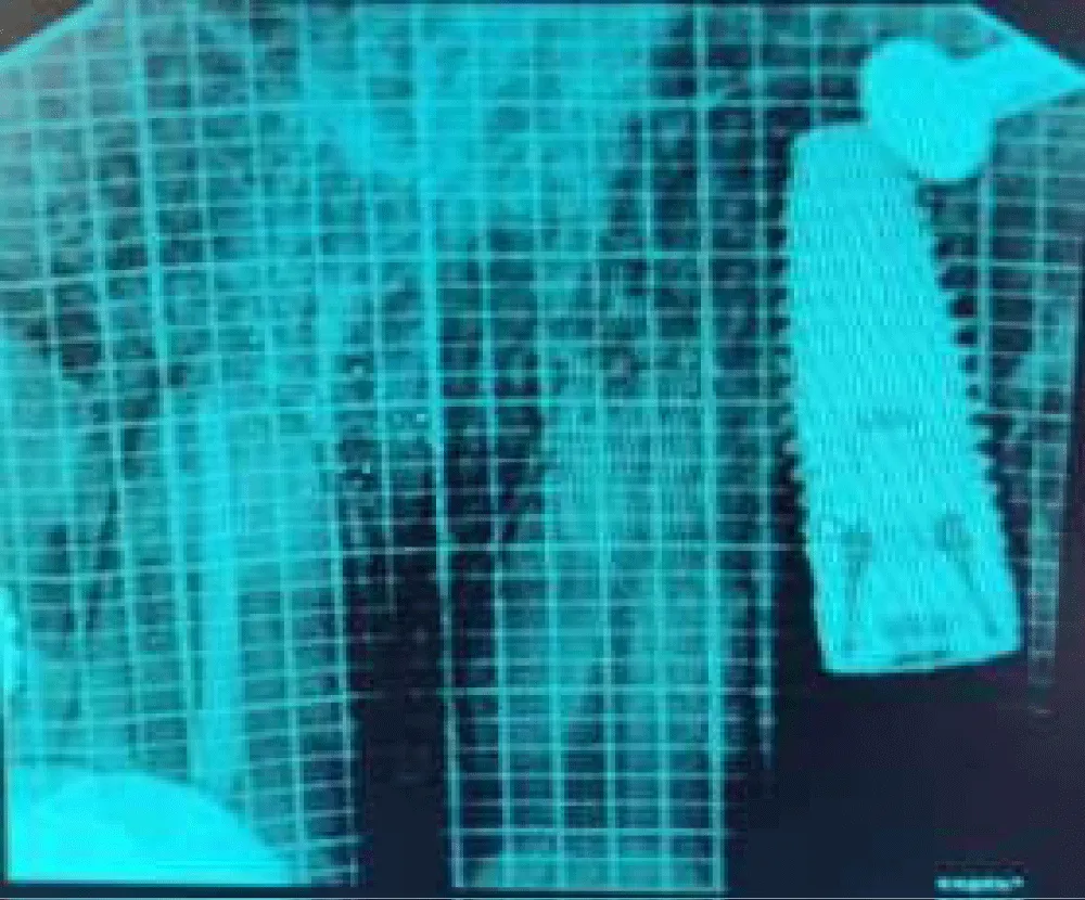
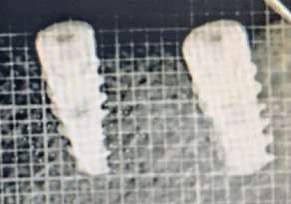
Bone gain was assessed using RVG and a grid with marking of 1mm2. Marginal bone level was measured by measuring the interproximal height of bone, which is defined as the distance measured between the apical ends of the first thread of the implant and the most coronal point of the interproximal crestal bone. This value was recorded using the RVG with Grid. This parameter was recorded at baseline and 3 months postoperatively. Measurements were taken by counting the number of gridlines between 2 specific points in the radiograph taken with the grid, and drawing a straight line between two points by using the RVG software. The paralleling cone technique was used to standardise the radiograph. (Figures 5&8)
Sample size Estimation: The effect size of this study was determined as previously described. Since the study was to be conducted with 2 groups, it was determined that the number of individuals in each group should be at least n = 10, and a total of n = 20 individuals were required. The effect size level of the study was 1.35, a = 0.05 for significance, and calculated by an 81% power analysis.
The sample size estimation was performed at 5% alpha error (a = 0.05), with an effect size of 1.35 [Based on the findings from the previous literature, study done by Jwa Young Kim et al, 2014, with the mean difference in the Absolute volume of new bone at 8 weeks b/w groups] & the power of the study at 80%, revealed that a minimum of 20 samples was necessary for the present study.
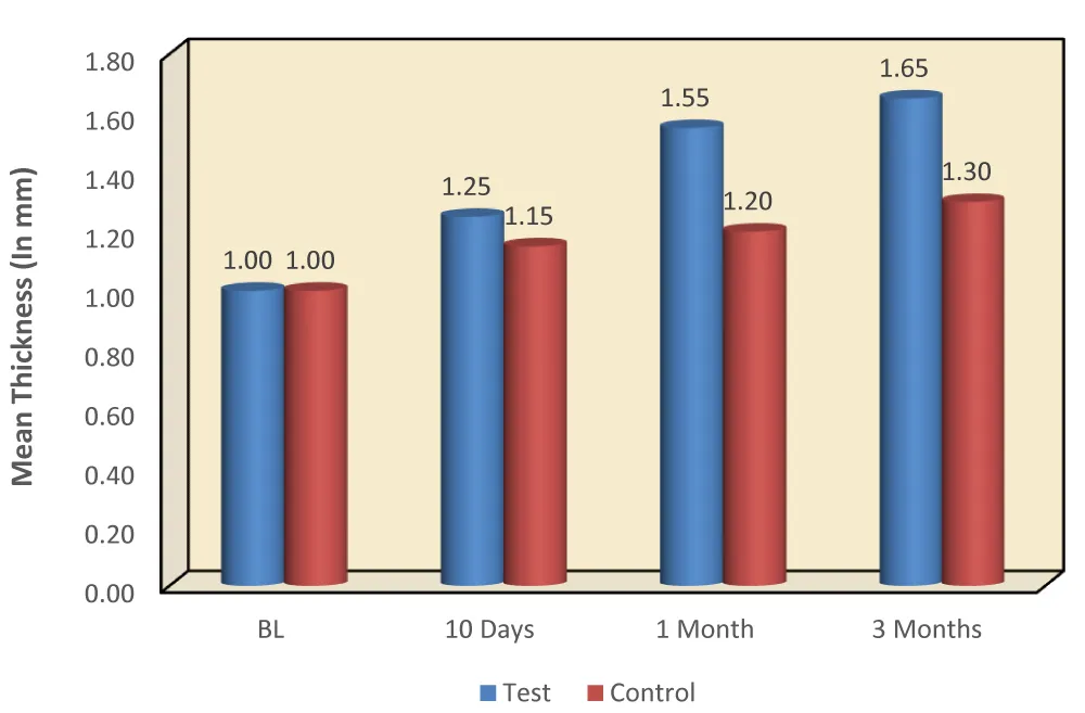
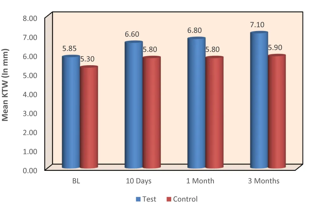
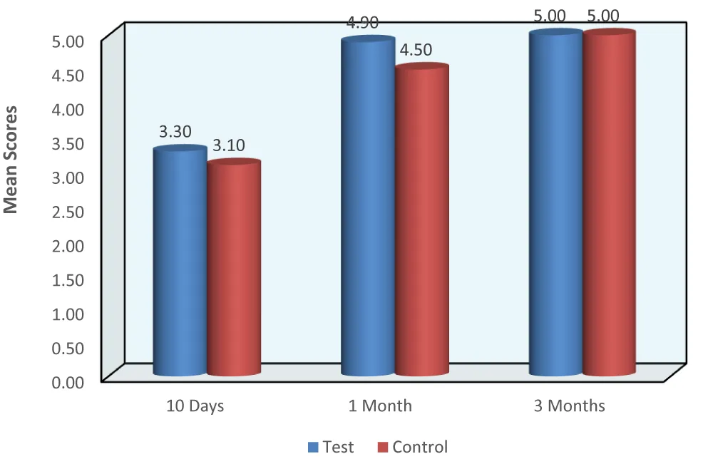
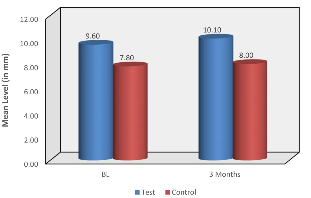
Statistical Analysis: Intragroup Comparison of mean Gingival Thickness (in mm) and mean Keratinised Tissue Width (in mm) b/w different time intervals in each group using Repeated Measures ANOVA Test. Multiple comparison of mean difference in Gingival Thickness (in mm) and mean difference in KTW b/w diff. time intervals in each group using Bonferroni’s post hoc Test. (Table 1) Comparison of mean Wound Healing scores b/w diff. time intervals in each group using Friedman’s Test followed by Wilcoxon Signed Rank post hoc Test. Comparison of mean Radiographic evaluation of Bone level (in mm) between Baseline and 3 Months period in each group using Wilcoxon Signed Rank Test. (Table 2) Intergroup comparison of mean Gingival Thickness and mean Keratinized Tissue Width (in mm) at different time intervals using Independent Student t Test. Comparison of mean Wound Healing Scores between 2 groups at 10th day, 1 month, and 3 Months period using Mann-Whitney Test. Comparison of mean Radiographic evaluation of Bone level (in mm) between 2 groups at Baseline and 3 Months period using the Mann-Whitney test. Graph (1-4).
Study design
Informed consent was obtained from all selected patients. Following the screening examination, eligible patients were enrolled in the trial. 12 male and 8 female patients were enrolled. All patients underwent routine scaling and root planing, and oral hygiene instructions were emphasised before being recalled for extraction and immediate implant placement. A detailed medical and dental history, along with blood investigations including complete blood count, was obtained before extraction. Gingival tissue thickness and keratinised tissue width were recorded before tooth extraction, and these measurements were considered as baseline values.
Pre-surgical Procedure
All patients were evaluated through detailed clinical history, clinical examination, and radiographic assessment. Phase I periodontal therapy was carried out for all patients. Gingival tissue thickness, keratinised tissue width, and Radiovisiography (RVG) with grid measurements were recorded at the predetermined immediate implant placement site.
Surgical procedure
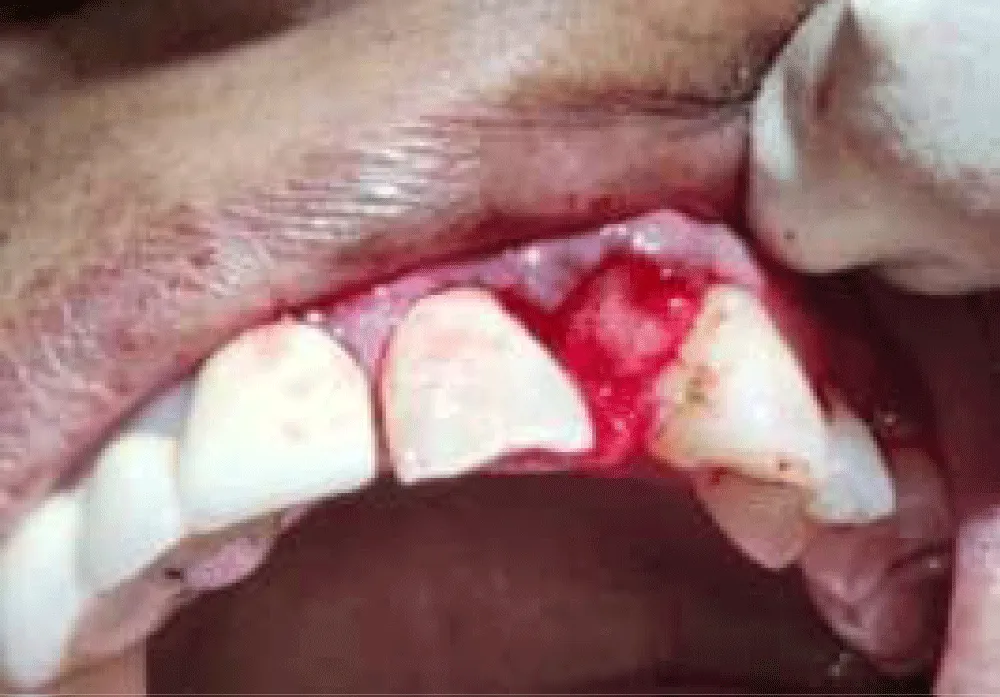
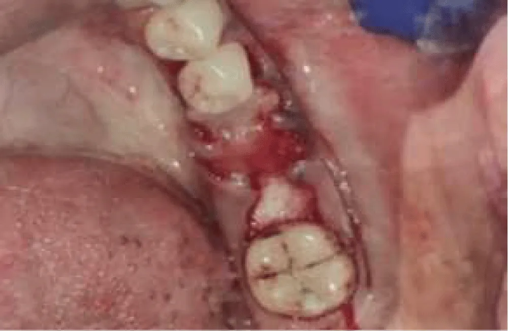
Pre-procedural mouth rinsing with chlorhexidine was performed before the procedure. Local anaesthesia was administered, followed by an incision made using a No. 15 blade, and a mucoperiosteal flap was elevated with a periosteal elevator. Thorough debridement of inflamed granulation tissue was then carried out. Atraumatic tooth extraction was performed using a periotome, followed by sequential drilling with copious irrigation up to the desired dimensions, extending 2–3 mm apical to the socket apex to achieve adequate primary stability. The sterile implant package was then opened, and the implant was guided into position with light, steady finger pressure. Final implant installation up to the bone level was completed using a ratchet wrench. Cross-linked collagen membrane with or without silk fibroin was randomly allocated to sites with gingival thickness ≤1 mm. (Figures 4 and 7). The flaps were approximated and secured with sutures, followed by placement of a periodontal dressing.
Postoperative care
Postoperative instructions consisted of gargling with saline and betadine mouth rinse twice daily for 1 minute and avoiding brushing at the surgical site for 2 weeks. Postoperative pain and oedema were controlled using non-steroidal anti-inflammatory drugs (NSAIDs) and antibiotics. Patients were advised to consume only soft and warm food during the first week following surgery. After 10 days, the periodontal dressing and sutures were removed, and the surgical site was evaluated using the Landry, Turnbull, and Howley Healing Index, which was considered the baseline value. Patients were subsequently recalled for reinforcement of oral hygiene instructions and supragingival debridement as maintenance care. Clinical parameters for wound healing were recorded at the end of the 10th day, 1 Month, and 3 Months after the surgical procedure. Gingival tissue thickness and keratinised tissue width were assessed at 1 Month and 3Months following immediate implant placement. Radiographic changes were assessed at baseline and at 3Months using RVG with a grid marked at 1 mm² intervals.
Results
A total of 20 implant sites were included in the present randomised controlled clinical trial, of which 10 sites were allocated to the Test group (Cross-linked collagen membrane with silk fibroin), and 10 sites were allocated to the Control group (Cross-linked collagen membrane alone). The clinical parameters: gingival thickness and keratinised tissue width were recorded at baseline, 10 days, 1 month, and 3 months and wound healing was recorded at 10 days, 1 month, and 3 months. The radiographic parameter was recorded at baseline and 3 months. Scores obtained were subjected to statistical analysis.
Bonferroni’s post hoc Test was conducted to identify specific pairwise differences in gingival thickness and keratinised tissue width across different time points within each group, and Friedman’s Test was used to analyse wound healing scores across time intervals within each group, and Wilcoxon Signed Rank Test was performed to compare radiographic bone levels between baseline and three months within each group. Results are tabulated in Table I and Table II.
Discussion
This study is the first to comprehensively evaluate the clinical effects of a silk fibroin–incorporated collagen membrane on soft tissue augmentation around immediate implants. The present randomised controlled clinical trial evaluated and compared the clinical and radiographic outcomes of a cross-linked collagen membrane incorporating silk fibroin (test group) with a cross-linked collagen membrane alone (control group) at immediate implant sites over three months.
Gingival tissue thickness
A statistically significant increase in gingival thickness was observed in both groups over time. However, the test group demonstrated significantly greater gains at 1 month and 3 months compared to the control group.
The superior increase in gingival thickness observed in the test group may be related to the biological properties of silk fibroin. Altman and colleagues described silk fibroin as a biomaterial capable of supporting fibroblast adhesion and proliferation [8]. Vepari and Kaplan further demonstrated that silk-based scaffolds facilitate organised collagen deposition and maturation of the extracellular matrix [9].
In contrast to conventional collagen membranes that tend to degrade rapidly, silk fibroin exhibits controllable degradation characteristics. This prolonged structural presence may provide greater scaffold stability during the early phases of wound healing, thereby supporting the maintenance of soft tissue volume [9].
Previous studies by Tuna et al., Amy et al., and Nguyen et al. reported that degummed silk fibroin induces minimal inflammatory response and promotes favourable healing compared with certain synthetic biomaterials [10-12]. Furthermore, a review by Kundu et al. indicated that silk-based scaffolds can enhance vascular endothelial growth factor (VEGF) expression, thereby promoting neovascularisation, which plays a critical role in early soft tissue maturation [13].
Keratinised tissue width
Keratinised tissue width increased significantly in both groups; however, the test group demonstrated significantly greater gains at both evaluation intervals.
The greater increase in keratinised tissue width in the test group may be related to enhanced epithelial cell attachment and stratification facilitated by silk fibroin matrices. Additional factors may include improved epithelial–connective tissue interactions and stabilisation of the underlying connective tissue scaffold [14].
In vitro studies have demonstrated that silk fibroin supports keratinocyte adhesion and differentiation, potentially through integrin-mediated cellular signalling pathways [15]. Moreover, the mechanical integrity of silk fibroin helps maintain the regenerative space, allowing epithelial migration to occur over a stable connective tissue foundation [13].
Wound healing
Wound healing scores improved significantly in both groups throughout the study, with the test group exhibiting superior early healing at 1 month.
Zhang et al. reported that silk fibroin matrices can upregulate growth factors such as transforming growth factor-β (TGF-β) and vascular endothelial growth factor (VEGF), thereby supporting tissue regeneration [156]. Kundu et al. also observed improved neovascularisation and faster wound closure in silk-based scaffolds compared with conventional biomaterials [13].
Additionally, silk fibroin possesses considerable mechanical strength, which may reduce micromovement at the surgical site [13]. Mechanical stability during early healing is particularly important for predictable regenerative outcomes in implant therapy [17].
Naik et al., in a clinical case series evaluating a silk fibroin–collagen composite membrane in soft tissue defects, reported significant improvements in gingival thickness, keratinised tissue width, and wound healing in periodontal and implant-related defects at 1 and 3 months. These findings further support the potential benefits of silk-enhanced collagen membranes in soft tissue augmentation procedures [18].
Radiographic bone levels
Intergroup comparison did not reveal statistically significant differences in radiographic bone levels at three months. However, intragroup analysis showed significant improvement in the test group.
Preclinical studies have suggested that silk fibroin possesses osteoconductive and osteinductive properties [13]. Wang et al. demonstrated enhanced differentiation of mesenchymal stem cells toward an osteoblastic lineage when cultured on silk fibroin scaffolds [19]. Similarly, Kim et al. reported increased alkaline phosphatase activity and mineral deposition in silk-based constructs [20]. The combination of silk fibroin powder with platelet-rich fibrin (PRF) has also been reported to improve peri-implant tissue regeneration by providing a growth-factor-rich scaffold [21]. Furthermore, composites combining silk fibroin and collagen may integrate the biological activity of collagen with the mechanical strength of silk, potentially enhancing regenerative outcomes.
The absence of a statistically significant difference between groups at the 3-month interval may be explained by several factors, including the relatively short follow-up period, limited sample size, and the inherent limitations of radiographic methods in detecting early mineralisation changes. Bone remodelling is a gradual biological process, and longer observation periods may be required to identify definitive differences.
Despite promising results, the present study has certain limitations. Short follow-up duration (3 months), which may not capture long-term remodelling; two-dimensional radiographic evaluation, which may underestimate volumetric changes; lack of histologic assessment, which limits understanding of tissue quality; and absence of comparison with the gold standard connective tissue graft. Another limitation is that Patient-reported outcome measures (PROMs) such as postoperative pain, discomfort, and patient satisfaction were not evaluated, which are important parameters in contemporary implant research. Additionally, the study did not differentiate outcomes between anterior and posterior implant sites, although variations in soft tissue thickness, esthetic demands, and functional loading may influence clinical results in different regions of the mouth.
Given these limitations, future research should incorporate longer follow-ups, CBCT-based assessments, inclusion of PROMs, and site-specific evaluation of anterior and posterior implants and histologic evaluations to provide a comprehensive evaluation of tissue integration and regenerative efficacy.
Conclusion
This randomised controlled clinical trial shows the application of a cross-linked collagen membrane incorporating silk fibroin around immediate dental implants, demonstrating more favourable clinical and radiographic outcomes when compared with the use of a collagen membrane alone. The test group exhibited significant and sustained increases in gingival thickness and keratinised tissue width, along with more rapid and predictable wound healing. These observations suggest that the inclusion of silk fibroin may improve the peri-implant soft tissue response and facilitate early tissue stabilisation following immediate implant placement. Radiographic assessment also indicated better preservation of peri-implant bone levels in the test group, implying that silk fibroin may positively influence hard tissue adaptation and contribute to overall implant stability.
The consistent improvements observed in both soft tissue and hard tissue parameters highlight the potential role of silk fibroin as a valuable adjunct in regenerative implant therapy. Incorporating silk fibroin into cross-linked collagen membranes may therefore represent a promising approach to enhance healing outcomes and support the long-term success of immediate dental implant procedures.
- Titsinides S, Agrogiannis G, Karatzas T. Bone grafting materials in dentoalveolar reconstruction: a comprehensive review. Jpn Dent Sci Rev. 2019;55(1):26-32. Available from: https://doi.org/10.1016/j.jdsr.2018.09.003
- Pagni G, Pellegrini G, Giannobile WV, Rasperini G. Post-extraction alveolar ridge preservation: biological basis and treatments. Int J Dent. 2012;2012:151030. Available from: https://doi.org/10.1155/2012/151030
- Zubery Y, Nir E, Goldlust A. Ossification of a collagen membrane cross-linked by sugar: a human case series. J Periodontol. 2008;79(6):1101-7. Available from: https://doi.org/10.1902/jop.2008.070421
- Smidt A, Gutmacher Z, Sharon E. A novel collagen scaffold to simplify lateral augmentation of deficient ridges between natural teeth. Quintessence Int. 2019;50(7):576-82. Available from: https://doi.org/10.3290/j.qi.a42652
- Lee JH. Bone regeneration using silk fibroin membranes in critical calvarial defects. J Tissue Eng Regen Med. 2024;12(5):345-58.
- Lv Q, Hu K, Feng Q, Cui F. Fibroin/collagen hybrid hydrogels with crosslinking method: preparation, properties, and cytocompatibility. J Biomed Mater Res A. 2008;84(1):198-207. Available from: https://doi.org/10.1002/jbm.a.31366
- Jiang S, Yu Z, Zhang L, Wang G, Dai X, Lian X, et al. Effects of different aperture-sized type I collagen/silk fibroin scaffolds on the proliferation and differentiation of human dental pulp cells. Regen Biomater. 2021;8:rbab028. Available from: https://doi.org/10.1093/rb/rbab028
- Altman GH, Diaz F, Jakuba C, Calabro T, Horan RL, Chen J, et al. Silk-based biomaterials. Biomaterials. 2003;24(3):401-16. Available from: https://doi.org/10.1016/s0142-9612(02)00353-8
- Vepari C, Kaplan DL. Silk as a biomaterial. Prog Polym Sci. 2007;32(8-9):991-1007. Available from: https://doi.org/10.1016/j.progpolymsci.2007.05.013
- Thurber AE, Omenetto FG, Kaplan DL. In vivo bioresponses to silk proteins. Biomaterials. 2015;71:145-57. Available from: https://doi.org/10.1016/j.biomaterials.2015.08.039
- Nguyen TP, Nguyen QV, Nguyen VH, Le TH, Huynh VQN, Vo DVN, et al. Silk fibroin-based biomaterials for biomedical applications: a review. Polymers (Basel). 2019;11(12):1933. Available from: https://doi.org/10.3390/polym11121933
- Yucel T, Lovett ML, Kaplan DL. Silk-based biomaterials for sustained drug delivery. J Control Release. 2014;190:381-97. Available from:
- Kundu B, Rajkhowa R, Kundu SC, Wang X. Silk fibroin biomaterials for tissue regeneration. Adv Drug Deliv Rev. 2013;65(4):457-70. Available from: https://doi.org/10.1016/j.addr.2012.09.043
- Strenge JT, Smeets R, Nemati F, Fuest S, Rhode SC, Stuermer EK. Biodegradable silk fibroin matrices for wound closure in a human 3D ex vivo approach. Materials (Basel). 2024;17(12):3004.
- Min BM, Lee G, Kim SH, Nam YS, Lee TS, Park WH. Electrospinning of silk fibroin nanofibers and its effect on the adhesion and spreading of normal human keratinocytes and fibroblasts in vitro. Biomaterials. 2004;25(7-8):1289-97. Available from: https://doi.org/10.1016/j.biomaterials.2003.08.045
- Zhang HM, Yang ML, Xi JZ, Yang GY, Wu QN. Mesenchymal stem cells-based drug delivery systems for diabetic foot ulcer: a review. World J Diabetes. 2023;14(11):1585-602. Available from: https://doi.org/10.4239/wjd.v14.i11.1585
- Retzepi M, Donos N. Guided bone regeneration: biological principle and therapeutic applications. Clin Oral Implants Res. 2010;21(6):567-76. Available from: https://doi.org/10.1111/j.1600-0501.2010.01922.x
- Davies JE. Understanding peri-implant endosseous healing. J Dent Educ. 2003;67(8):932-49. Available from: https://pubmed.ncbi.nlm.nih.gov/12959168/
- Naik J, Kumar A, Sharma R. Collagen and silk fibroin membranes in tissue regeneration: a clinical case series. J Indian Soc Periodontol. 2024;28(3):312-8.
- Wang Y, Rudym DD, Walsh A, Abrahamsen L, Kim HJ, Kim HS, et al. In vivo degradation of three-dimensional silk fibroin scaffolds. Biomaterials. 2008;29(24-25):3415-28. Available from: https://doi.org/10.1016/j.biomaterials.2008.05.002
- Jang ES, Park JW, Kweon H, Lee KG, Kang SW, Baek DH, et al. Restoration of peri-implant defects in immediate implant installations by Choukroun platelet-rich fibrin and silk fibroin powder combination graft. Oral Surg Oral Med Oral Pathol Oral Radiol Endod. 2010;109(6):831-6. Available from: https://doi.org/10.1016/j.tripleo.2009.10.038

 Save to Mendeley
Article Alerts
Subscribe to our articles alerts and stay tuned.
 This work is licensed under a Creative Commons Attribution 4.0 International License.