Open J Biol Sci
Characteristics and outcome of fetal nuchal translucency above 3.5 mm in the first trimester
Cátia Lourenço1*, Inês Gouveia1, Ana Carriço2 and Francisco Valente1
2Department of Pediatric Cardiology, Vila Nova de Gaia / Espinho Hospital Center, Portugal
Cite this as
Lourenço C, Gouveia I, Carriço A, Valente F (2019) Characteristics and outcome of fetal nuchal translucency above 3.5 mm in the first trimester. Open J Biol Sci 4(1): 003-006. DOI: 10.17352/ojbs.000013Objective: To describe the natural history of fetuses with a nuchal translucency (NT) above 3.5 mm when crown-rump length measures between 45 and 84 mm.
Methods: We performed a retrospective cohort study of fetuses with first trimester NT above 3.5 mm between January 2013 and March 2017.
Results: A total of 75 cases with NT above 3.5 mm in the first trimester were identified. 3 cases were excluded (lost to follow-up), so that 72 cases were included. There were additional first trimester markers of aneuploidy in 16 cases and 5 cases of major structural abnormalities diagnosed in the first trimester ultrasound. 2 cases declined invasive testing. The karyotype was abnormal in 30 cases (43%), including 17 cases of trisomy 21. There were 25 terminations of pregnancy (34.7%), 11 fetal deaths (15.3%) and 36 livebirths (50%). The 36 live-born infants were followed. In this group 3 cases of trisomy 21, 1 case of unilateral hypoplasia of orbicularis oris, 1 Noonan-like syndrome, 1 case of 8p23.1 duplication (benign variant) and 1 case of lower limb lymphedema were observed.
Conclusion: The prognosis of fetal NT above 3.5 mm in the first trimester is poor when associated with karyotype abnormalities or structural abnormalities but is substantially better if neither of these conditions is observed.
Introduction
First trimester scan screens for major fetal abnormalities and chromosomal abnormalities early in pregnancy. Increased nuchal translucency (NT) between 11 weeks and 13 weeks plus 6 days is associated with abnormal karyotype, structural malformations, fetal death and disorders with late postnatal onset, such as Noonan’s syndrome.1-3 Many published studies have suggested that increased NT is associated with poor fetal outcome. The long-term prognosis of these infants is poorly documented [1-3].
The aim of this study was to determine the course of pregnancy and neonatal outcome of fetuses with NT above 3.5 mm diagnosed between 11 and 13 weeks plus 6 days and the prognosis of live-born infants.
Materials and Methods
This retrospective study was carried out between January 2013 and March 2017 in a single center. All cases with NT above 3.5 mm between 11 and 13 weeks +6 days were included in the study using data from our ultrasound base (Astraia). Additional data were retrieved from patients’ files and collected in a database. The General Electric Voluson E8 and Voluson 730 Expert were used, with a 4-8 MHz transabdominal probe (and transvaginal probe, when needed), for ultrasound.
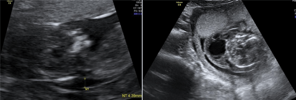
The diagnosis of fetal NT above 3.5 mm was made in the midsagittal sonographic views of the fetal neck and according to the criteria of the Fetal Medicine Foundation. The presence of other first trimester markers of aneuploidy was noted and fetal anatomy was evaluated. When this diagnosis was made, the parents received counselling explaining the key points of management based on natural history: its association with genetic abnormalities, cardiac malformations and others, the possibility of spontaneous resolution specially if isolated, and the risk of intra-uterine fetal death. The patient is offered fetal genetic testing (karyotype and cGH array if the first is normal) by chorionic villus sampling at 11-14 weeks or amniocentesis from 16 weeks onwards and early fetal echocardiography. Ongoing pregnancies were monitored by means of regular sonography until delivery. The outcomes assessed were abnormal karyotype, major structural anomaly, fetal death, termination of pregnancy (TOP), live birth and/or neonatal death. An abnormal karyotype was defined as a karyotype other than 46 XX or 46 XY by cells obtained from chorionic villus sampling or amniocentesis. Major structural anomaly was defined as a structural malformation identified by ultrasound, echocardiography or post-natal evaluation that would be life-threatening, result in long term disability or negatively impact individual outcome. Autopsy was proposed for all intrauterine and neonatal deaths and termination of pregnancy. Outcome was defined as normal if the pregnancy resulted in a liveborn neonate with no abnormalities detectable in the newborn period.
Results
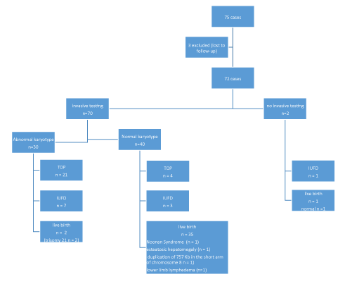
Between January 2013 and March 2017, 75 cases of NT above 3.5 mm were diagnosed between 11 and 13 weeks plus 6 days gestation (Figure 1). 3 cases were excluded because follow-up pf the pregnancy was not possible. 72 cases were included in the study. Maternal and pregnancy characteristics are presented in table 1.
Mean maternal age at diagnosis was 33.5 years. The mean size of NT at diagnosis was 5,3 mm. The parents were routinely offered fetal karyotyping. 2 cases refused invasive testing. There were 69 cases were chorionic villus sampling (CVS) was performed and 5 cases were amniocentesis was performed (in which 4 cases also performed CVS). The karyotype was abnormal in 30 cases (42.9%). The chromosomal abnormalities were as follows: trisomy 21 in 17 cases (24%); Turner’s syndrome in 4 cases (5,7%); trisomy 18 in 3 cases (4,3%); trisomy 13 in 2 cases (1,4%), and other chromosomal abnormalities in 4 cases (5,7%) (Table 2). In 16 cases (22.2%) there were additional first trimester ultrasound markers of aneuploidy, namely absent nasal bone, reversed a-wave of the ductus venosus and tricuspid regurgitation. There were no differences in mean NT between the two groups. (Table 3). In 5 cases (6.9%) increased NT was associated with fetal hydrops. In 9 cases (12.5%) there were structural anomalies diagnosed by ultrasound or echocardiography in the first trimester. In these 9 cases there were 6 cases of abnormal karyotype and 3 cases of a normal karyotype (Table 4). There were no differences in mean NT between the group with malformations and without them (Table 5).
Of those fetuses with a normal karyotype (40 cases) there were 7 cases were cGH array was not available. Of the remaining 33 cases with normal karyotype, there was one case with a gain of material in 10 q, not described in the literature. The study of the pregnant woman revealed a similar result to that of the fetus.
There was persistence of increased nuchal fold in the second trimester in 5 cases (6.94%).
There were 11 cases (15.3%) of intra-uterine fetal death, all before 17 weeks. There were 3 cases of normal karyotype and 1 case on unknown karyotype (the patient declined invasive testing). In these 4 cases the autopsy revealed minor anomalies in 1 case, a truncus arteriosus in 1 case, a common atrioventricular valve and a polymalformative syndrome in 1 case and fetal growth restriction in 1 case. (Table 6)
There were 25 cases (34.7%) of termination of pregnancy before 24 weeks gestation: 14 cases of trisomy 21, 3 cases of 45 X0, 3 cases of trisomy 18, 4 cases of normal karyotype (associated with major structural abnormalities) and 1 case of trisomy 13 (Table 7).
Of the 36 live-born infants (50%) there was one case of unilateral agenesis of orbicularis oris, 1 case of clinical Noonan Syndrome (associated with neurodevelopment delay), 2 cases of trisomy 21 (1 with atrioventricular septal defect), 1 case of esteatosic hepatomegaly (surveillance in metabolic disease appointment), 1 case of duplication of 757 Kb in the short arm of chromosome 8 (this variant is classified as benign) and 1 case of lower limb lymphedema. The remaining live born had no malformations and normal development. Preterm birth occurred in 5 of the 50 liveborn children (10%), 4 were late preterm and 1 at 32 weeks (Figure 2).
Discussion and Conclusion
This study represents a cohort of fetuses with first-trimester NT above 3.5 mm and evaluates the incidence, characteristics and outcomes of fetuses with this diagnosis. We evaluated the rates of abnormal karyotype, fetal abnormality, intra-uterine fetal death, termination of pregnancy and normal development of the newborn infants because we considered this information useful to appropriately counselling parents in a prenatal diagnosis setting and for decision-making in these situations [1,4].
There is a strong association of increased nuchal translucency and fetal aneuploidy, ranging from 29 to 60% in the literature [5,6]. Trisomy 21 is the most frequent aneuploidy in our study, which is in accordance with the majority of the literature [5,7]. Lore et all found that the prevalence of Turner syndrome is higher than trisomy 21 in their study [8,9].
The pregnancy outcome in strongly related to the presence of structural abnormalities and/or genetic abnormalities, so that the literature points out the need for subsequent evaluation after the detection of an increased nuchal translucency or cystic hygroma. In this group, karyotype and array cGH, detailed anatomic assessment, and fetal echocardiography are essential components of subsequent evaluation [4].
Our findings demonstrate a strong association of increasing nuchal translucency thickness in the first trimester with high rates of abnormal karyotype, major congenital anomaly, intra-uterine fetal death and abnormal outcome among fetuses with cystic hygroma, as depicted in figure 2. A recent meta-analysis showed an additional yield of microarray is 5 % in fetuses with a normal karyotype and increased NT, highlighting its diagnostic and prognostic importance [8]. In our study there was only a case in which the cGH array revealed a 10Q duplication, mas the study of the parents showed the same duplication in the mother. This could be due to small sample size.
The paediatric prognosis of euploid fetuses with increased NT depends largely on disorders with late postnatal onset. However, in our population there was a good prognosis in liveborn children with normal karyotype and no prenatal diagnosis of malformations.
Our study shows limitations. It concerns a small sample size and it is a retrospective cohort study. Additionally no distinction was made between septated and non-septated cases and there are some papers showing that visualization of nuchal septations during first-trimester genetic screening is a powerful risk factor for chromosomal anomalies, independent of increased NT [10]. Microarray analysis was not available for all cases and so submicroscopic pathogenic copy number variants may have been missed.
- Graesslin O, Derniaux E, Alanio E, Gaillard D, Vitry F, et al. (2007) Characteristics and outcome of fetal cystic hygroma diagnosed in the first trimester. Acta Obstet Gynecol Scand 86: 1442-1146. Link: http://bit.ly/2Y9ObsZ
- Lajeunesse C, Stadler A, Trombert B, Varlet MN, Patural H, et al. (2014) Firsttrimester cystic hygroma: prenatal diagnosis and fetal outcome. J Gynecol Obstet Biol Reprod (Paris) 43: 455-462. Link: http://bit.ly/2M6lkUm
- Rosati P, Guariglia L (2000) Prognostic value of ultrasound findings of fetal cystic hygroma detected in early pregnancy by transvaginal sonography. Ultrasound Obstet Gynecol 16: 245-250. Link: http://bit.ly/32LhdmC
- Scholl J, Durfee SM, Russell MA, Heard AJ, Iyer C, et al. (2012) First-trimester cystic hygroma: relationship of nuchal translucency thickness and outcomes. Obstet Gynecol 120: 551-559. Link: http://bit.ly/32GrY9z
- Malone FD, Ball RH, Nyberg DH (2005) First-trimester septated cystic hygroma: prevalence, natural history and pediatric outcome. Obstet Gynecol 106. Link: http://bit.ly/2GkHUoo
- Kharrat R, Yamamoto M, Roume J, Couderc S, Vialard F, et al. (2006) Karyotype and outcome of fetuses diagnosed with cystic hygroma in the first trimester in relation to nuchal translucency thickness. Prenat Diagn 26: 369–372. Link: http://bit.ly/2xZmIQb
- Scholl J, Durfee SM, Russell MA, Heard AJ, Ecker J, et al. (2012) First-Trimester Cystic Hygroma : Relationship of Nuchal Translucency Thickness and Outcomes. Obstet Gynecol 120: 551-559. Link: http://bit.ly/32GrY9z
- Grande M, Jansen FAR, Blumenfeld YJ, Fisher A, Odibo AO, et al. (2015) Genomic microarray in fetuses with increased nuchal translucency and normal karyotype: A systematic review and meta-analysis. Ultrasound Obstet Gynecol 46: 650–658. Link: http://bit.ly/32JwYKL
- Lore S, Lore L, Luc DC, Dominique VS, Koenraad D, et al. (2018) First trimester cystic hygroma colli: retrospective analysis in a tertiary center. Eur J Obstet Gynecol Reprod Biol 231: 60-64. Link: http://bit.ly/2M6nVgQ
- Mack LM, Lee W, Mastrobattista JM, Belfort MA, Van den Veyver IB, et al. (2017) Are First Trimester Nuchal Septations Independent Risk Factors for Chromosomal Anomalies? J Ultrasound Med 36: 155-161. Link: http://bit.ly/32KgRfW
Article Alerts
Subscribe to our articles alerts and stay tuned.
 This work is licensed under a Creative Commons Attribution 4.0 International License.


 Save to Mendeley
