Global Journal of Biotechnology and Biomaterial Science
Biological toxicity evaluation of traditional medicine white tea extract liquid
Rui Zhang1#, Mingyue Lin1$, Chenxin Wang1, Yufan Li1, Xinggang Zhao2, Jun Liu2, Li Chen3* and Qin Zou1*
2Shenzhen Weiduo Technology Co., Ltd. Shenzhen 518000, China
3Analytical & Testing Center, Sichuan University, Chengdu 610064, China
#These authors contributed equally to this work
Li Chen, Analytical & Testing Center, Sichuan University, Chengdu 610064, P.R. China, E-mail: [email protected]
Cite this as
Zhang R, Lin M, Wang C, Li Y, Zhao X, et al. (2023) Biological toxicity evaluation of traditional medicine white tea extract liquid. Glob J Biotechnol Biomater Sci 9(1): 001-008. DOI: 10.17352/gjbbs.000018Copyright Licence
© 2023 Zhang R, et al. This is an open-access article distributed under the terms of the Creative Commons Attribution License, which permits unrestricted use, distribution, and reproduction in any medium, provided the original author and source are credited.Purpose: The aim of this study was to evaluate the safety and toxicity of intravenous use of White Tea Extract Liquid (WT) in rabbits through routine blood tests and biochemical and histopathological means.
Design and methods: This study used the WT product produced by the company and the New Zealand rabbits at 2.0~2.5 kg of weight. Rabbits were divided into two groups: Control and WT groups. In the WT group, the diluted WT injection volume needed to be injected was calculated according to their body weight on days 1, 4, 7 and 11...... for 4 weeks respectively. During the1, 2 and 4 weeks of the experiment, blood routine, blood biochemistry and the histopathological sections of the heart, spleen, lung, trachea, kidney, bladder, uterus, testicle, stomach, liver, small intestine, big intestine were evaluated in the study.
Findings: By comparison, the blood routine test did not deviate from the normal ranges provided by the diagnostic laboratory. The blood biochemical indexes of all experimental rabbits are within a reasonable range and did not detect clinically significant abnormalities between treatment and control groups. The WT had no obvious inflammatory change in the whole tissue and no harmful effect on the organs of various systems of the rabbits’ system for 4 weeks.
Conclusions: After intravenous injection for 4 weeks in rabbits, white tea herb atomization liquid did not produce obvious toxic effects on the animals.
Introduction
Commercially available technologies to transform a liquid dosage form into an aerosol for medical inhalation purposes have evolved significantly over the last century. As a noninvasive and easy drug administration route, drug delivery via aerosolization administration also has the potential to minimize toxicity without compromising efficacy and improve patient compliance [1]. Traditional Chinese medicine combined with aerosolization technology is different from traditional smelling inhalation and smoke inhalation, forming a new drug delivery mode. It has the characteristics of small atomized particles, easy absorption and a high drug utilization rate. It has been widely used in the treatment of respiratory, blood circulation and other system diseases, with remarkable efficacy [2].
White tea herbal atomization liquid (WT) is a traditional Chinese herbal liquid product with white tea extract and glycerol as the main components. Its main functions are to clear away heat and detoxify, and have significant anti-inflammatory effects [3]. The extract of white tea contains several polyphenolic compounds belonging to the flavan-3-ol (catechin) family that possesses a wide range of biological activities, including anticancer, antiviral and antibacterial effects [4]. Glycerol has been widely used in food, medicine, and other industries. The existing experiment proved that rats had nasally inhaled glycerol for 90 days and were exposed to a dose of 750 mg/kg through the analysis and comparison of body weight and food consumption, blood, organs, etc, which had not produced obvious toxic effects on rats [5].
The way of atomization inhalation of drugs will lead to a large difference in the dose of each aerosol, thus greatly reducing the final bioavailability of herbal liquid in the specific experimental process [6]. In order to find a suitable way to test the safety and toxicity of Chinese herbal atomizing liquid, we chose to inject the sample into the New Zealand rabbits through auricular intravenous injection and conducted a biological evaluation on experimental rabbits regularly. Compared with atomization inhalation, intravenous injection is easy to operate and can control the amount of administration. At the 1st, 2nd, and 4th weeks after the administration, the clinical symptoms and food intake of the animals were observed, and their blood routine, blood biochemistry, and organ histopathology were examined.
Materials and methods
Materials
White tea flavor stock solution, Shenzhen Weiduo Technology Co., Ltd (Guangdong, China). Disposable vacuum blood collection tubes were purchased from Yongkang Medical Products Co., Ltd (Shandong, China). The Hematoxylin and Eosin (H&E) staining kit was purchased from Beyotime Biotechnology Co., Ltd (Jiangsu China). All chemicals in the experiments were in Analytical Reagent (AR) grade and used without further purification.
Design
Rabbits’ weighing and intravenous injection volume calculation: In a sterile environment, the WT was diluted from 1200 mg/ml to 30 mg/ml(C), and physiological saline was taken as the solution. On injection days, each animal received a single marginal ear vein dose of a diluted WT at a dose of 10 mg/kg body weight (AD). The choice of the dose was based on published work [7] indicating that this is a therapeutic dose in most animal species (mammals and not) for all routes of administration. The animals were weighed(M)and the injection dose was calculated by Equation 1 as:
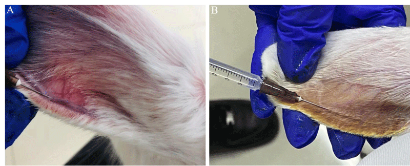
10(male:5 and female:5) New Zealand white rabbits were routinely fed for 2 weeks before the experiment, and the body weight after feeding was 2.3-2.7 kg. The animals were randomly assigned to two separate surgical groups (an injection group (n = 6, male:2 and female:2) and a no injection normal group (n = 4; male:2 and female:2). All the procedures in this animal experiment were approved by the Ethics Committee of West China Hospital at Sichuan University. Before the injection of the sample, the hair on the vein of the ear margin was shaved and the injection site was cleaned with iodine disinfection (Figure 1A). 1ml bore needle syringes were used to obtain better precision. After injection (Figure 1B), apply pressure on the application site for 5 - 10 seconds to stop bleeding. The sample was injected on the day, 1, 3, 5 and 7…... of feeding and the control group did not receive any drug injection. The mental state, hair color, diet, feces and body weight of rabbits were observed.
Complete blood count tests: At the 1st, 2nd, and 4th weeks after intravenous injection, one female and one male rabbit were randomly selected from the injection group to be anesthetized by intravenous injection of sodium pentobarbital at a dose of 40 mg/kg at the ear margin. After blood was collected in an EDTA anticoagulant tube, it was gently mixed, and then the blood routine parameters were measured on the automatic animal blood cell analyzer (BC-2800 Vet, Mindray, China). The routine blood tests contain 18 items, which are WBC (White Blood Cell), Lymph# (Lymphocytes Number), Mon# (Monocytes Number), Gran# (Granulocytes Number), Lymph% (Lymphocytes Percentage), Mon% (Monocytes Percentage), Gran% (Granulocytes Percentage), RBC (Red Cell Count), HGB (Hemoglobin), HCT (Hematocrit), MCV (Mean Red Cell Volume), MCH (Mean Red Cell Hemoglobin), MCHC (Mean Red Cell Hemoglobin Content), RDW(Red Cell Distribution Width), PLT (Platelet), MPV (Mean Platelet Volume), PDW (Platelet Distribution Width) and PCT (Procalcitonin). Statistical analysis was carried out using the mean ± standard deviation (SD).
Blood biochemical test: After the blood was collected with the blood collection vessel without anticoagulant, it was centrifuged at 3000r / min for 10min after standing for 0.5h and the upper serum was taken and the blood biochemical indexes were measured with the automatic animal biochemical analyzer (BS-240 Vet, Mindray, China). The biochemical blood tests contain 20 items, which are Fe (Iron), Mg (Magnesium), AST (Aspartate Aminotransferase), ALT (Alanine Aminotransferase), ALP (Alkaline Phosphatase), ALB (Albumin), γ-GT (Gamma-Glutamyl), Glu (Glucose), UA (Uric Acid), Ca (Calcium), P (Phosphorus), CK (Creatine Kinase), LDH (Lactate Dehydrogenase), UREA (Urea), CREA (Creatinine), TC (Total Cholesterol), TG (Triglyceride), TP (Total Protein), T-bil (Total Bilirubin) and GLO (Globulin). Statistical analysis was carried out using the mean ± standard deviation (SD).
HE staining of organs: At the 1st, 2nd and 4th weeks after intravenous injection, one female and one male rabbit were randomly selected from the injection group for euthanasia, and the heart, spleen, lung, trachea, kidney, bladder, uterus, testicle, stomach, liver, small intestine and big intestine were quickly removed and fixed with 4% paraformaldehyde for more than 24 hours. Samples for histological analysis were embedded in paraffin wax. The cutting sections (5 μm in thickness) were stained with H&E and observed by microscope (Ti-U, Nikon, Japan).
Results
The general state of animals
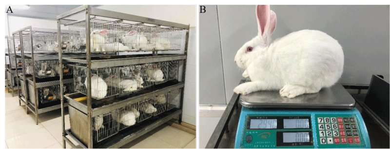
All the animals survived until the end of the study and no perioperative period complications were encountered. The rabbits in each group were in a good mental state, and there were no obvious abnormalities in their hair color, behavioral activities, food intake, drinking water and fecal respiration (Figure 2A). The body weight of the test animals was measured before the start of the test and at the 1st, 2nd, and 4th weeks after the injection of WT (Figure 2B). The body weight of the test rabbits increased from 2.44 ± 0.06 to 2.94 ± 0.14. There was no significant increase or decrease in the body weight of the animals in each male and female group, and there was no statistical difference. It was seen that long-term consumption did not produce any significant change in growth or food ingestion, as Espinosa et al. previously reported [8]. In addition, research by Francisca et al. showed that White tea extract consumption did not significantly change body weight gain, food intake, food efficiency, protein efficiency ratio, apparent protein digestibility, nitrogen balance, or the apparent absorption of calcium, phosphorus, magnesium and zinc [9].
Effects of WT on the blood routine of rabbits
Complete blood counts are not only important basic indicators for biomedical evaluation but also the change of its results can reveal many diseases as well [10]. They can help to determine the overall physiological state of the experimental animals. WBC, Mon, Mon%, Gran, Gran%, Lymph%, and Lymph can reflect the susceptibility of animals, suggesting inflammation and infection; RBC, HGB, HCT, MCV, MCH, MCHC, and RDW can reflect whether the animal is anemia; and PLT, MPV, PDW, and PCT can be used as one of the auxiliary diagnostic indicators for viral diseases, malignant tumors, and other diseases [11]. From the experimental results in Tables 1,2, it can see that the blood routine test did not deviate from the normal ranges provided by the diagnostic laboratory. Although several items were lower or higher than the reference ranges, the differences are relatively small. We consider these differences as minor variations from normal due to random variation within the herd and the detection of differences as a result of chance. The study of saral et al showed that white tea inhibited NF- κB-related inflammation, which inhibited its synthesis, leading to a significant decrease in the expression level of inflammatory factors such as IL-6 and TNF- α [12].
Effects of WT on the biochemical blood of rabbits
Blood biochemical examination is often used to detect the contents of various ions, sugars, lipids, proteins, various enzymes, hormones and various metabolites of the body in the blood [13]. It is a reflection of the functions and states of various tissues and organs of animals. Under normal health conditions, blood glucose (Glu) and inorganic salt ions (Fe, Mg, Ca and P) are stable in the body and kept in a certain balance range to maintain the normal metabolism and osmotic pressure of the body. Otherwise, glucose metabolism disorder and electrolyte imbalance will occur; [14]. AST, ALT and ALP are important indicators of liver function. When the liver cell membrane is damaged or cell necrosis, these enzymes will increase in the serum. By measuring the activity of enzymes in serum or plasma, the degree of liver cell damage can be reflected. Among them, because AST is mostly distributed in the myocardium, it will be released into the blood when the heart is damaged, causing the increase of AST, which is also an important indicator of heart function; UREA and CREA are indicators of kidney function. When various kidney diseases cause renal insufficiency, they will increase [15]. For the results of blood biochemical indexes analysis (Tables 3,4), no abnormalities were detected. By comparison, the blood biochemical indexes of all experimental rabbits are within a reasonable range and did not detect clinically significant abnormalities between WT and control groups. Tea and its components have been considered potential ingredients for ameliorating liver injuries in patients. In a double-blinded, placebo-controlled, randomized clinical trial, the green tea extract consumption (500 mg/day, 90 days) successfully decreased the levels of liver enzymes including ALT, AST and ALP in patients with non-alcoholic fatty liver disease [16]. In another clinical trial with hypercholesterolemic subjects, catechin-enriched green, and oolong tea treatments were found to significantly decrease body weight, BMI, fat, lipid peroxidation and lipid profiles (TG, TC, LDL-C, and HDL-C), while improving GSH, SOD, CAT, GPX and GR in the liver [17].
Tissue histopathological changes of rabbits’ main organs
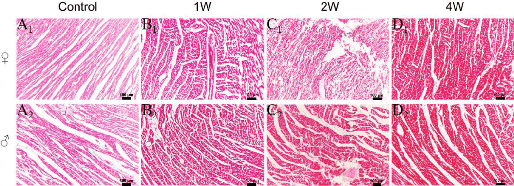
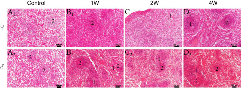
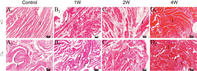
Circulatory system: The circulatory system is a continuous and closed pipeline system, including the cardiovascular system and lymphatic systems. As the power pump of the cardiovascular system, the heart can promote the continuous circulation of blood in the blood vessels and provide sufficient blood flow, oxygen, and various nutrients for organs and tissues [18]. HE staining of myocardial tissue showed that the muscle fiber structure of the rabbit myocardial tissue in the control groups (Figure 3A1, A2) and the experimental groups (Figure 3B1-D2) was clear, and the transverse striations were visible. The ladder-like disc structure was seen at the junction of the myocardial fibers, and no inflammatory cell infiltration was found. As a part of the lymphatic system, the spleen acts as a hematopoietic organ in the embryonic period. After the bone marrow starts to make blood, the spleen has evolved into the largest lymphoid organ in the human body, with the functions of hematopoietic stem cells, antigen clearance, aging red blood cells, and immune response [19]. On the fresh section of the spleen in the experimental group (Figure 4B1-D2), most of the tissues were crimson red pulp, with abundant blood vessels distributed there. Various fine structures formed by the lymphoid tissue in the spleen were regularly distributed along the blood vessels. Optical images of HE-stained sections showed that after week 4 of injection, no significant pathological changes were observed in the heart and spleen, indicating the biosafety of WT. Intake of tea and its extracts can decrease CVD risk, mainly by improving redox status, inhibiting inflammation, decreasing blood pressure, ameliorating hyperlipidemia, regulating endothelial function, preventing myocardial damage, and regulating sympathetic nerve activity [20]. This further explains the protective effect of tea on the cardiovascular system.
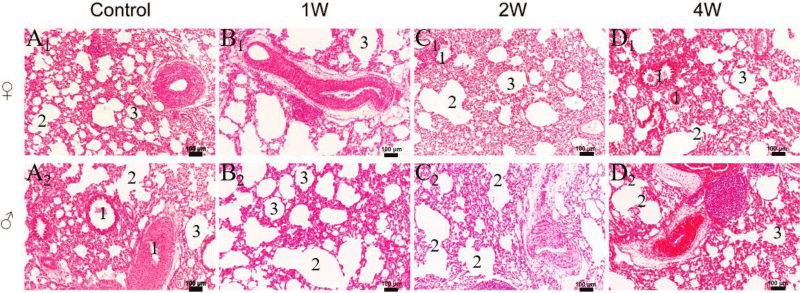
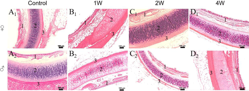
Respiratory system: The main function of the lungs is to exchange air. The lungs are rich in blood vessels. The alveoli exchange gas with the blood through the respiratory membrane (i.e., the alveolar-capillary membrane, also known as the blood-air barrier) [21]. It can be seen from the HE staining images of the control group (Figure 5A1, A2) and the WT group (Figure 5B1-D2) that the lung tissue structure was clear and complete. The alveolar structure was normal, the alveolar wall is smooth, and the bronchi are evenly distributed. There were no edema and inflammatory cells in the surrounding tissues. The trachea and bronchus are the air channels outside the lung. The wall of the trachea is divided into mucosa, submucosa and adventitia from the inside to the outside [22]. The mucosa is composed of abundant ciliated cells, goblet cells and basal cells, which have the function of clearing foreign bodies, bacteria, and other harmful substances. The complete and clear mucosa, submucosa and adventitia parts can be observed in the control group (Figure 6A1, A2) and sample groups (Figure 6B1, D2). The submucosa contained abundant blood vessels, lymphoid tissues, and many mixed tracheal glands. The structure of tissue had no significant change and the results provided evidence of the safety of white tea on the respiratory system. Acute lung injury and acute respiratory distress syndrome are serious clinical complications with high morbidity and mortality [23]. A study in mice showed that treatment with tea catechins improved histopathological lesions, reduced pro-inflammatory markers, and improved lung regeneration capacity by improving the expression of Ki67, PCNA and Ang-1 proteins [24].
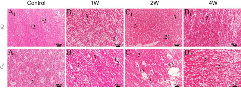
Urinary system: To study the effect of the sample on the urinary system of rabbits, we observed the kidney and bladder sections of rabbits injected with WT at different times. The kidney can maintain the water and electrolyte balance of the body by regulating the urine production process and changing the excretion of water and inorganic ions [25]. In the normal control group (Figure 7A1, A2), the glomeruli of the kidney were full, there was no gap with the capsule wall, and the structure was neat. In the WT group (Figure 7B1-D2), both renal corpuscle, and renal tubule can be observed. There was no obvious expansion or atrophy of glomeruli, which showed similar conditions to the control group. The bladder is an organ that stores urine. Its muscle layer is thick, and the mucosa contains many folds. When the bladder is filled, the folds decrease or disappear. From the sections of experimental groups (Figure 8B1-D2), we can see that the bladder at this time was in an empty state, and the epithelium had multiple layers of epithelial cells connected without damage. These findings showed that intravenous injection of WT for nebulization for 4 weeks did not cause histopathological changes in the urinary system in rabbits. So far, it has been reported that tea catechins improve albuminuria in diabetic nephropathy in a human trial [26]. The main mechanism involved the activation of DGKα, so protecting kidney activity. It has been shown that tea catechins could act also as an iron chelator, resulting in decreased ROS production and subsequent protection against kidney damage [27].
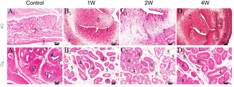
Reproductive system: The uterus and testis are important components of the reproductive system. By observing the changes in the sections of these two organs, we can further study the effects of WT on the physiological activity and reproductive offspring of rabbits. From the sections of the uterus (Figure 9B1-D1), the perimetrium, myometrium, and endometrium of the injected sample groups were demarcated. The endometrial mucosa formed many branched folds that protruded into the official cavity. The official cavity was irregular and contained abundant blood vessels, uterine glands and a large number of stromal cells. Testicular sections (Figure 9B2-D2) showed that the testicular capsule tissue was composed of dense connective tissue rich in elastic fibers with uniform thickness, and the seminiferous tubules were closely arranged which contained abundant germ cells and Sertoli cells. There was no abnormality in the interstitial tissue. Therefore, the white tea had no obvious inflammatory change in the whole tissue and no harmful effect on the rabbits’ reproductive system for 4 weeks. tea catechins can stimulate and regulate the hormones that govern the menstrual cycle. They can also modulate the levels of progesterone and oestrogenic hormones, as well as improve fertility, and are a possible treatment for increasing the pregnancy rate [28]. Testicular tissue is predisposed to the action of free radicals due to its high rate of cell division. Thanks to the antioxidant properties of tea, inflammation and DNA fragmentation can be reduced, and sperm motility and viability can be increased [28,29].
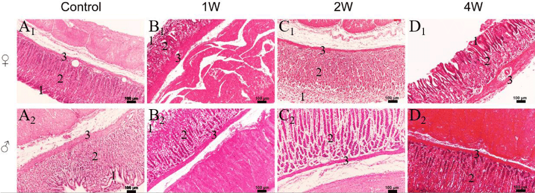
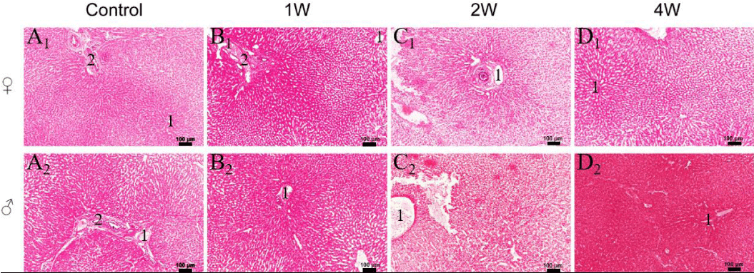
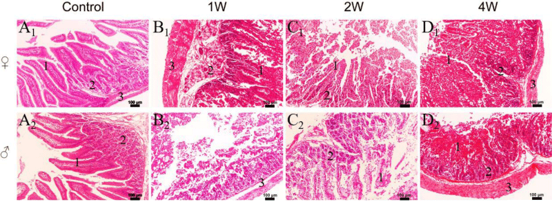
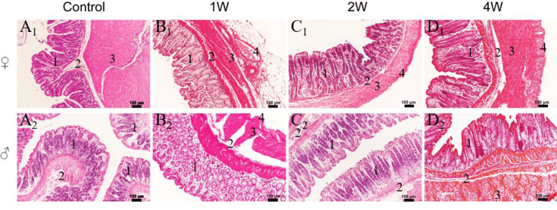
Digestive system: We also take HE-stained images of other organs of the digestive system, which included the stomach, liver, small intestine, and large intestine. Gastric sections in the control groups (Figure 10A1, A2) and WT groups (Figure 10B1-D2) showed tight junctions of mucus cells with important protective effects in the gastric fovea, and no goblet cells were found. In addition, many fundus glands were closely arranged, containing lots of blue chief cells and red parietal cells. In the liver section (Figure 11A1-D2), the hepatocytes were arranged radially around the central vein to form a plate-like structure and the hepatic cords were connected to form a network channel. Also, no noticeable inflammatory cells are captured. Both the small intestine (Figure 12B1-D2) and the large intestine (Figure 13B1-D2) had complete and clear mucosa, submucosa and muscular in the WT groups. The small intestine and large intestine glands were densely and regularly arranged, containing a lot of absorptive cells and goblet cells, as well as a small number of stem cells and endocrine cells. On the whole, the structure of organ tissue had no significant change and a few inflammatory cells were found. Thus, a 4-week injection of WT was safe for the rabbits’ digestive system. Tea extracts and their bioactive components possess strong anti-inflammatory activity, thus can be a potential agent for arthritis, sepsis and SLE, with the mechanisms mainly including the regulation of pro-inflammatory and anti-inflammatory factors, like interleukins, chemokines, TNF-α, NF-κB, and COX-2, as well as the related signaling pathways [30]. Inflammation of the intestinal epithelial barrier is important in diseases such as inflammatory bowel disease or Crohn’s disorder. Rodent models have shown that catechin-rich white tea extract improves the integrity of the intestinal barrier [31]. This generates a modulation in the translocation of endotoxins derived from the intestine and their consequent pro-inflammatory responses [32]. Tea catechins treatment significantly increased antioxidant capacity and reduced the signs of related colitis, as well as mitigating less severe symptoms white tea extract also reduces the entry of immunoreactive nitrotyrosine into the colon and reduces the over-regulation of intercellular adhesion molecule 1 (ICAM-1) [33,34].
Conclusion
After the rabbits were given WT intravenously for 1, 2, and 4 weeks, our findings suggested that there were no biological and toxicological changes related to herbal solution in animal body weight, hematology, blood biochemistry and organ HE sections in each experimental group. In conclusion, the WT may not cause significant adverse effects on rabbits. Finally, all studies on WT have been conducted in cells or animal culture models. Further clinical trials are needed to provide conclusive evidence of the protective effects of the biological toxicity evaluation.
Funding
This work was supported by the Biological Evaluation of Atomizer and Herbal Atomizing Liquid [No.21H0588] Effect of Atomized Liquid of Traditional Chinese Medicine on Metabolism and Ultrastructure of Stem Cells [No.21H0928]Innovation Reform Project of Postgraduate Education of Sichuan University in 2021, and Experimental Technology Research Project of Sichuan University [SCU221099].
- Li C, Liu S, Luo G, Wang G, Zhang B, Nie Q. Comparison of plasma pharmacokinetics of Tanreqing solution between intratracheal aerosolization and intravenous injection in rats. Biomed Chromatogr. 2018 Mar;32(3). doi: 10.1002/bmc.4116. Epub 2017 Nov 22. PMID: 29027677.
- Dugas HL, Peters JI, Williams RO 3rd. Nebulization of mycophenolate mofetil inhalation suspension in rats: comparison with oral and pulmonary administration of Cellcept®. Int J Pharm. 2013 Jan 30;441(1-2):19-29. doi: 10.1016/j.ijpharm.2012.12.016. Epub 2012 Dec 20. PMID: 23262419.
- Granato D, Prado-Silva Ld, Alvarenga VO, Zielinski AAF, Bataglion GA, Morais DRd, Sant'Ana AdS. Characterization of binary and ternary mixtures of green, white and black tea extracts by electrospray ionization mass spectrometry and modeling of their in vitro antibacterial activity. LWT - Food Science and Technology. 2016; 65: 414-420.http://dx.doi.org/10.1016/j.lwt.2015.08.037
- Almajano MP, Carbó R, Jiménez JAL, Gordon MH. Antioxidant and antimicrobial activities of tea infusions. Food Chemistry. 2008; 108(1):55-63. http://dx.doi.org/10.1016/j.foodchem.2007.10.040
- Peace MR, Mulder HA, Baird TR, Butler KE, Friedrich AK, Stone JW, Turner JBM, Poklis A, Poklis JL. Evaluation of Nicotine and the Components of e-Liquids Generated from e-Cigarette Aerosols. J Anal Toxicol. 2018 Oct 1;42(8):537-543. doi: 10.1093/jat/bky056. PMID: 30371842; PMCID: PMC6203128.
- Mulder HA, Patterson JL, Halquist MS, Kosmider L, Turner JBM, Poklis JL, Poklis A, Peace MR. The Effect of Electronic Cigarette User Modifications and E-liquid Adulteration on the Particle Size Profile of an Aerosolized Product. Sci Rep. 2019 Jul 15;9(1):10221. doi: 10.1038/s41598-019-46387-2. Erratum in: Sci Rep. 2020 Mar 13;10(1):4975. PMID: 31308389; PMCID: PMC6629610.
- Nair AB, Jacob S. A simple practice guide for dose conversion between animals and human. J Basic Clin Pharm. 2016 Mar;7(2):27-31. doi: 10.4103/0976-0105.177703. PMID: 27057123; PMCID: PMC4804402.
- Espinosa Ruiz C, Cabrera L, López-Jiménez JÁ, Zamora S, Pérez-Llamas F. Effects of long-term ingestion of white tea on oxidation produced by aging and acute oxidative damage in rats. J Physiol Biochem. 2018 Feb;74(1):171-177. doi: 10.1007/s13105-017-0591-z. Epub 2017 Sep 19. PMID: 28929453.
- Pérez-Llamas F, González D, Cabrera L, Espinosa C, López JA, Larqué E, Almajano MP, Zamora S. White tea consumption slightly reduces iron absorption but not growth, food efficiency, protein utilization, or calcium, phosphorus, magnesium, and zinc absorption in rats. J Physiol Biochem. 2011 Sep;67(3):331-7. doi: 10.1007/s13105-011-0079-1. Epub 2011 Feb 19. PMID: 21336649.
- Abdel-Hamid TM, Farahat MH. Effect of dietary mannan-oligosaccharides on some blood biochemical, haematological parameters and carcass traits in purebred New Zealand White and crossbred rabbits. Animal Production Science. 2016; 56(12). http://dx.doi.org/10.1071/an15032
- Sembratowicz I, Zięba G, Cholewinska E, Czech A. Effect of Dietary Flaxseed Oil Supplementation on the Redox Status, Haematological and Biochemical Parameters of Horses' Blood. Animals (Basel). 2020 Nov 30;10(12):2244. doi: 10.3390/ani10122244. PMID: 33265987; PMCID: PMC7760300.
- Saral S, Dokumacioglu E, Mercantepe T, Atak M, Cinar S, Saral O, Yildiz L, Iskender H, Tumkaya L. The effect of white tea on serum TNF-α/NF-κB and immunohistochemical parameters in cisplatin-related renal dysfunction in female rats. Biomed Pharmacother. 2019 Apr;112:108604. doi: 10.1016/j.biopha.2019.108604. Epub 2019 Feb 22. PMID: 30798139.
- Williams LB, Co C, Koenig JB, Tse C, Lindsay E, Koch TG. Response to Intravenous Allogeneic Equine Cord Blood-Derived Mesenchymal Stromal Cells Administered from Chilled or Frozen State in Serum and Protein-Free Media. Front Vet Sci. 2016 Jul 22;3:56. doi: 10.3389/fvets.2016.00056. PMID: 27500136; PMCID: PMC4956649.
- Jeklova E, Leva L, Knotigova P, Faldyna M. Age-related changes in selected haematology parameters in rabbits. Res Vet Sci. 2009 Jun;86(3):525-8. doi: 10.1016/j.rvsc.2008.10.007. Epub 2008 Nov 28. PMID: 19041105.
- Sun L, Gao Y, Zhang W, Liu H, Sun D. Effect of high fluoride and high fat on serum lipid levels and oxidative stress in rabbits. Environ Toxicol Pharmacol. 2014 Nov;38(3):1000-6. doi: 10.1016/j.etap.2014.10.010. Epub 2014 Nov 5. PMID: 25461561.
- Tsai YJ, Chen BH. Preparation of catechin extracts and nanoemulsions from green tea leaf waste and their inhibition effect on prostate cancer cell PC-3. Int J Nanomedicine. 2016 May 6;11:1907-26. doi: 10.2147/IJN.S103759. PMID: 27226712; PMCID: PMC4866752.
- Venkatakrishnan K , Chiu HF , Cheng JC , Chang YH , Lu YY , Han YC , Shen YC , Tsai KS , Wang CK . Comparative studies on the hypolipidemic, antioxidant and hepatoprotective activities of catechin-enriched green and oolong tea in a double-blind clinical trial. Food Funct. 2018 Feb 21;9(2):1205-1213. doi: 10.1039/c7fo01449j. PMID: 29384173.
- Meki MH, Miller JM, Mohamed TMA. Heart Slices to Model Cardiac Physiology. Front Pharmacol. 2021 Feb 4;12:617922. doi: 10.3389/fphar.2021.617922. PMID: 33613292; PMCID: PMC7890402.
- Kallendrusch S, Merz F, Bechmann I, Mayr SG, Zink M. Long-Term Tissue Culture of Adult Brain and Spleen Slices on Nanostructured Scaffolds. Adv Healthc Mater. 2017 May;6(9). doi: 10.1002/adhm.201601336. Epub 2017 Feb 20. PMID: 28218503.
- Ueshima H. Explanation for the Japanese paradox: prevention of increase in coronary heart disease and reduction in stroke. J Atheroscler Thromb. 2007 Dec;14(6):278-86. doi: 10.5551/jat.e529. Epub 2007 Dec 17. PMID: 18174657.
- Gao P, Zhao Z, Zhang C, Wang C, Long K, Guo L, Li B. The therapeutic effects of traditional Chinese medicine Fusu agent in LPS-induced acute lung injury model rats. Drug Des Devel Ther. 2018 Nov 13;12:3867-3878. doi: 10.2147/DDDT.S181798. PMID: 30518997; PMCID: PMC6239118.
- Han MN, Kim JH, Choi SH. Evaluation of Biomechanical Properties and Morphometric Structures of the Trachea in Pigs and Rabbits. In Vivo. 2022 Jul-Aug;36(4):1718-1725. doi: 10.21873/invivo.12884. PMID: 35738586; PMCID: PMC9301417.
- Unno K, Furushima D, Nomura Y, Yamada H, Iguchi K, Taguchi K, Suzuki T, Ozeki M, Nakamura Y. Antidepressant Effect of Shaded White Leaf Tea Containing High Levels of Caffeine and Amino Acids. Molecules. 2020 Aug 3;25(15):3550. doi: 10.3390/molecules25153550. PMID: 32756488; PMCID: PMC7435365.
- Almatroodi SA, Almatroudi A, Alsahli MA, Aljasir MA, Syed MA, Rahmani AH. Epigallocatechin-3-Gallate (EGCG), an Active Compound of Green Tea Attenuates Acute Lung Injury Regulating Macrophage Polarization and Krüpple-Like-Factor 4 (KLF4) Expression. Molecules. 2020 Jun 20;25(12):2853. doi: 10.3390/molecules25122853. PMID: 32575718; PMCID: PMC7356789.
- van Furth LA, Leuvenink HGD, Seras L, de Graaf IAM, Olinga P, van Leeuwen LLJT. Exploring Porcine Precision-Cut Kidney Slices as a Model for Transplant-Related Ischemia-Reperfusion Injury. 2022.
- Bao H, Peng A. The Green Tea Polyphenol(-)-epigallocatechin-3-gallate and its beneficial roles in chronic kidney disease. J Transl Int Med. 2016 Sep 1;4(3):99-103. doi: 10.1515/jtim-2016-0031. Epub 2016 Sep 23. PMID: 28191529; PMCID: PMC5290885.
- Liu L, Liu B, Li J, Zhen S, Ye Z, Cheng M, Liu W. Responses of Different Cancer Cells to White Tea Aqueous Extract. J Food Sci. 2018 Oct;83(10):2593-2601. doi: 10.1111/1750-3841.14351. Epub 2018 Sep 25. PMID: 30251345.
- Hinojosa-Nogueira D, Pérez-Burillo S, Pastoriza de la Cueva S, Rufián-Henares JÁ. Green and white teas as health-promoting foods. Food Funct. 2021 May 11;12(9):3799-3819. doi: 10.1039/d1fo00261a. PMID: 33977999.
- Tang GY, Meng X, Gan RY, Zhao CN, Liu Q, Feng YB, Li S, Wei XL, Atanasov AG, Corke H, Li HB. Health Functions and Related Molecular Mechanisms of Tea Components: An Update Review. Int J Mol Sci. 2019 Dec 8;20(24):6196. doi: 10.3390/ijms20246196. PMID: 31817990; PMCID: PMC6941079.
- Liu L, Wu X, Zhang B, Yang W, Li D, Dong Y, Yin Y, Chen Q. Protective effects of tea polyphenols on exhaustive exercise-induced fatigue, inflammation and tissue damage. Food Nutr Res. 2017 Jun 1;61(1):1333390. doi: 10.1080/16546628.2017.1333390. PMID: 28659745; PMCID: PMC5475289.
- Rahman SU, Li Y, Huang Y, Zhu L, Feng S, Wu J, Wang X. Treatment of inflammatory bowel disease via green tea polyphenols: possible application and protective approaches. Inflammopharmacology. 2018 Apr;26(2):319-330. doi: 10.1007/s10787-018-0462-4. Epub 2018 Mar 12. PMID: 29532213.
- Luo K, Ma C, Xing S, An Y, Feng J, Dang H, Huang W, Qiao L, Cheng J, Xie L. White tea and its active polyphenols lower cholesterol through reduction of very-low-density lipoprotein production and induction of LDLR expression. Biomed Pharmacother. 2020 Jul;127:110146. doi: 10.1016/j.biopha.2020.110146. Epub 2020 Apr 22. PMID: 32334376.
- Farzaei MH, Bahramsoltani R, Abbasabadi Z, Braidy N, Nabavi SM. Role of green tea catechins in prevention of age-related cognitive decline: Pharmacological targets and clinical perspective. J Cell Physiol. 2019 Mar;234(3):2447-2459. doi: 10.1002/jcp.27289. Epub 2018 Sep 6. PMID: 30187490.
- Truong VL, Jeong WS. Cellular Defensive Mechanisms of Tea Polyphenols: Structure-Activity Relationship. Int J Mol Sci. 2021 Aug 24;22(17):9109. doi: 10.3390/ijms22179109. PMID: 34502017; PMCID: PMC8430757.

Article Alerts
Subscribe to our articles alerts and stay tuned.
 This work is licensed under a Creative Commons Attribution 4.0 International License.

 Save to Mendeley
